Heel Pain? That Was Achilles’ Problem As Well…

The death of Achilles was not mentioned in Homer‘s Iliad, but appeared in later Greek and Roman poetry and drama concerning events after the Iliad, such as later in the Trojan War. Here and in the myths surrounding the war, Achilles died from a heel wound as the result of an arrow—possibly poisoned—shot by Paris.

According to a myth, Achilles’ mother dipped the infant Achilles in the river Styx, holding onto him by his heel, thus he became invulnerable where the waters touched him — that is, everywhere but the areas covered by her thumb and forefinger.

This is a statue of Achilles dying at the Corfu Achilleion. (www.wikipedia.com)

Achilles Tears, Ruptures and Tendinitis:

The achilles tendon is one of the largest and strongest tendons in the body.  The gastrocnemius, soleus and plantaris muscles all combine to form this structure which inserts onto the back of the heel bone (calcaneous), see image below.  The achilles tendon and its’ muscles cross three joints, the knee, ankle, and subtalar joint.  Uniquely, it has no tendon sheath like other tendons.  Supposedly the achilles tendon has poor blood supply 2-6 cm above its’ insertion point making it vulnerable to rupture, degeneration and other variations of mechanical failure, however, this is a much debated point.  Some studies have shown that the vascular supply is quite good in this region, the fact remains that this is the location of most achilles tendon ruptures/tears/degeneration.  Other very common problems usually include calcification of the tendon at it’s insertion into the calcaneous creating spurs and loose bony bodies.  Often a large spur can develop near the insertion of the achilles that can place painful forces against the tendon itself (Haglund’s Deformity).

(Image Source)

Prevention:

Muscles, tendons and ligaments weaken, shorten and become ineffectual structures if exercise and activity are forgotten.  The office person with feet shoved under a desk for 8 hours a day with toes down and achilles tendon slowly shortening who then runs out on the weekend to do some high impact activity is a prime candidate for achilles problems.  People who use cowboy boots, high heels or other shoes with a higher heel also may suffer the same fate if they transition rapidly to shoes without a heel.  If the achilles is repeatedly injured and left untreated, the chronic inflammatory process may eventually result in degeneration of the tendon, calcification and pain only repairable with surgery.   Stretching for 30 minutes a day and warm ups before exercising are both great preventative medicine.    Take your time warming up for an event, don’t rush things.   A large review of studies recently demonstrated that stretching and warm up exercises did help prevent injury but the evidence was not overwhelming.

(image source)

Diagnoses:

In the office, a large tear or rupture can be felt as a hole or defect in the normally solid and smooth achilles tendon.  Chronic tendinitis that has turned into degeneration of the tendon may leave an enlarged and/or calcified portion of the tendon that can be felt and seen visibly bulging under the skin.

Achilles tendinitis presents as chronic pain that worsens with activity and is relieved with rest.

For ruptures and tears, the normal story usually involves the patient relating an audible pop, feelings of being hit with a baseball bat in the heel, a fall from significant height, and any variation of “weekend warrior” stories.  Like any tissue of the body, “if not used, it becomes easily abused”.

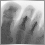
Imaging such as an MRI (performed in a Hospital) or Ultrasound (performed in our office) can shed light on the seriousness of the injury, helping to differentiate between tears, ruptures and chronic tendinitis.

Often the patients story helps in the diagnosis.  Was there an acute event such as a fall and a popping sound or has the pain been gradually worsening over the last few months?  A recent event that was the beginning of the pain indicates rupture or tear whereas chronic soreness would indicate more of an ongoing tendinitis (inflammation of the tendon) or tendinosis (degeneration of the tendon).  Imaging is normally a good idea because one type of problem often masquerades as another muddled by the fact that as patients, we don’t often remember specific details months after a problem began.

Treatment:

For the problems of tendinitis and tendinosis the usual suspects are called upon: rest, ice, NSAIDS (see post: Do I Have Plantar Fasciitis?).  The achilles tendon absorbs enormous amounts of  of tensions and energy when walking and running, this means that taking the tension off the tendon while it heals is paramount.  Often a podiatrist will prescribe cessation of running and walking for a number of weeks or at least cutting down the amount significantly.  The foot in question will be placed in a ski boot looking device (CAM walker) for bad cases.  For less severe cases, to protect the achilles tendon from its normal motions we will often incorporate a heel lift to take more tension off the tendon.  These boots, called CAM walkers, can be removed so the pt can follow a protocol of simple stretches and range of motion exercises as their healing progresses.


(image source)

Depending on the seriousness of the injury , within 3-4 or more weeks the pt should begin eccentric exercises.  A concentric exercise would be curling a dumbbell.  Eccentric exercises are just the opposite of shortening muscles, it’s allowing the muscle to lengthen slowly while resisting the movement.  It is unclear why this is so important, but studies have been very clear that this is a necessary and effective way of treating tendinitis and similar injuries.

Massage is a great way to loosen up the tendon and relieve pain, the video below mentions rollers as an easy way to accomplish this.

Here is a fantastic video Dr. Metzl has put together explaining the in’s and out’s of achilles tendinitis.   (www.runnersworld.com/injury-prevention-recovery/inside-doctors-office-keep-achilles-tendinitis-away?cm_mmc=NL-TrainingExtra-_-1174884-_-01222013-_-Inside-the-Doctor%27s-Office%3A-Achilles-Tendinitis)

Grastym/Astym, Topaz Microdebridement, Shockwave Therapy and Tenex have all had success with treating tendinitis/tendinosis pain and encouraging healing (see post: I Think I Have Plantar Fasciitis).

Partial and complete tears are a little more tricky.  Obviously the ends of the torn ligaments need to be brought back together or at least held together to allow for them to heal.  This either means surgically suturing the pieces together with possible reinforcement coming from cadaver (donated) tissues or synthetic tissues, or serial casting/CAM Boot therapy allowing the ligaments to be brought back together by the body’s own natural healing processes.

If you want to begin a battle, state boldly on the internet that one (surgery or casting) is superior than another.  The evidence shows that within the first year of surgical intervention there are lower rates of rerupture but not in the long term.  Surgical correction was shown to have more complications such as infection.  The casting/conservative approach had more reruptures in the first year but fewer complications.  Both were quite similar in outcomes after a year or more of recovery.  I’m not going to take sides here as treatment protocols are completely specific to a patients situation, but something tells me that surgery may be the better option for a “younger” healthy patient who can keep the wound clean post op.

Achilles tendon calcification/spurring normally requires surgical resection of the offending portion of the tendon with reattachment of the remaining tendon to the calcaneous.  The amount of rehab time depends on how invasive the surgery had to be to remove the spur/calcifications.

In general, you are looking at wearing a cast/boot for 6-8 weeks or so with a gradual return to activities.  Physical therapy is usually prescribed for strengthening and stretching guidance.  Full sporting involvement should be delayed for 6 months after the surgery or from the beginning of casting protocols.  This of course depends on the patient, the severity of the tear and so on.

So if the weekend calls with some court, trail, floor or wall with your name on it, take your time warming up, if it’s been a long while, take it easy, you’ll be happier without six months of rehab.   🙂

This is an evolving area of surgery and medicine with many new products and procedures being attempted at this time.  Before committing to surgery or some other treatment program make sure you do your own reading or get a second opinion if you have any  doubts at all.

Feel free to send any questions our way.

Dan Preece, DPM


Salt Lake Podiatry Center P-LLC

Dan Preece, DPM  &  Darren Groberg, DPM

Office: 801-532-1822, Fax: 801-532-7544
Address: 144 South 700 East SLC, UT 84102-1109
                
     Foot & Ankle Specialists
Posted in Achilles Tendon | Tagged , , , , , , , , , , | Leave a comment

Do I Have Plantar Fasciitis?

Plantar Fasciitis (PF)?  What is it?

On the foot of the average person there exists a very thick band of tissue that extends from the bottom of the heel bone (calcaneous) to the ball of the foot, inserting into the skin and tissues around each of the toes.  This tissue spans the arch of the foot like a string on a bow.  With each step, as weight is placed on the ball of the foot, the arch tends to flatten a little (or a lot depending on the person) tightening this band.  Too much tension on the plantar fascia causes micro tears and a painful inflammatory response.

(image source)

An increase in weight, change in activity such as a new running routine, long hours on your feet, poor shoe gear, tight calf muscles etc. can all contribute to the problem.  Also, when you sleep, your feet tend to point downward, this allows the calf muscles and plantar fascia to tighten up during the night.  This can also happen after sitting down for a few hours.  When you begin walking the shortened tissues are stretched out causing that “first steps” pain.

When the plantar fascia is injured it can enter into a perpetual state of inflammation and degeneration.  This process can be controlled but if the patient continues to walk through the pain the body never gets a chance to heal itself.  The process repeats itself many times, scar tissue begins to build up and local nerves may become entrapped or permanently irritated and enlarged.  This is the time when the patient should begin to contemplate surgery.  If conservative treatments fail for several months with no improvement being appreciated, then more invasive measures should be taken.  Thankfully, before this level of pathology is reached, the majority of patients find great relief with the conservative measures discussed below if diagnosed correctly in time.

Diagnoses of PF:

PF is normally diagnosed based on the patients story, an x-ray and quick physical exam.  The patient’s feet, specifically the heels, hurt with the first steps taken in the morning or after any prolonged period of rest.  The pain gradually improves with activity but may persist throughout the day worsening after each period of prolonged rest.  Pain is usually found if you press deeply into center of the bottom of the heel.

The plantar fascia can be visualized with ultrasound, x-ray and MRI.  Thickening of the PF as well as evidence of inflammation are all pieces of evidence used in diagnoses of PF.  Partial tears can sometimes be seen but are not normally appreciated.

I was told I have heel spurs…

Do heel spurs equal pain or a diagnoses of PF??  NO!!  In the past it was once taught in medical schools that having heel spurs equated to heel pain.  Studies have since demonstrated that many people have spurring at the heel without any symptoms whatsoever.  The key is this: if it doesn’t hurt don’t fix it, even if you find a huge spur on x-ray.

(image source)

How is PF Treated?

Conservative treatment is the first line of attack.

Stretching routines should focus on the plantar fascia, achilles tendon and calf muscles.  A number of good studies have shown that sitting with your leg bent into your lap and stretching your toes backwards is the most effective stretch for PF (see picture below).  Typical runner’s stretches are best with the foot flat on the ground and the body leaning towards a wall alternating straight and bent knee positions (see pictures below).  Hold the stretching position for greater than 40 seconds at each try and repeat for 10-15 minutes per day.  An even better approach is one 15 min session in the morning and and one at night especially if calf muscle tightness is thought to be the culprit.  There are several devices to help in your stretching routine: Thera-Band, Icing, heat packs, rest are also useful.  Some patients swear by night splints.  Night splints hold the foot at 90 degrees from the leg keeping the plantar fascia and calf muscles stretched out all through the night helping to prevent morning first steps pain and diminishing pain throughout the day.


(image source)


Supportive shoes are the baseline of treatment.  These should have a firm cup around the back of the heel, little give when the shoe is twisted lengthwise and only bend at the ball of the foot.  No particular brand of shoe is always a slam dunk, find one that meets these criteria and wear them around the store to verify comfort.  This being said, Asics, New Balance and Brooks have a fairly good track record.   Your feet are more important than the cost of the shoes.  However, more expensive shoes don’t necessarily mean a better fit or function.


Orthotics have proven to be quite effect for some individuals (much discussion about orthotics in future posts to come).  The current debate is whether $300+ custom orthotics or $30-50 over the counter orthotics are the better treatment.  Recent research has shown that custom orthotics perform better in energy expenditure efficiency and pain relief.  A good fitting orthotic supports the foot, reducing excessive movement such as pronation and supports the foot and plantar fascia allowing increased healing.  Our experience shows better outcomes are had by having custom orthotics made for your specific arch shape compared with buying over the counter products that you may not fit well.  Custom orthotics from our lab routinely last over 10 years while over the counter orthotics may only last a few months.

Custom orthotics fabricated on our office typically last over 10 years and quickly pay for themselves when compared with the cost of supplying yourself with over the counter orthotics for that period of time.   We guarantee that our orthotics will bring you satisfactory pain relief or return them for a full refund.  The cost of the orthotics includes fabrication, casting, fitting and modifications.

(image source)

NSAIDS are a class of drug that moderate the inflammatory process of the body responsible for swelling and pain, both of which are part of the healing process.  The danger with these drugs can be their affects on unhealthy kidneys, increased chance of bleeding and effects on the stomach and intestine contributing to ulcerations.   NSAIDS should not be taken for periods longer than a few months and therefore are only an adjunct treatment option for PF pain.

Steroid injections into the plantar fascia are usually enough to knock out the pain for 2-3 months if not for years at time.   Excessive steroid injections can lead to weakening (atrophy) of the tendons, skin, fat pads and capsules of the foot near the injection site.  Most podiatrists will not inject a patient more than 3-4 times per year (this is a topic of great debate, and each podiatrist may give you a different story).  I have seen many cases of patients that need an injection every few years and are “good to go” for quite a long time.  Others don’t experience as much success.  When an injection is combined with supportive shoes, orthotics and stretching, pain will slowly start to diminish.

(image source)


Surgery: When is surgery finally the answer?  In the case of Plantar Fasciitis it is the last resort.  The traditional surgical approach is either total or partial release of the plantar fascia from it’s attachment to the calcaneous with possible removal of associated nerves.  This is either done through a larger open incision or through smaller endoscopic portals using a camera and instruments.  Results are quite encouraging with 80-95% of patients reporting successful outcomes.  The remaining 5-20% of patients may not experience pain relief at all, may have new pain elsewhere in the foot as joints realign, may develop infection and/or may experience damaged or trapped nerves.  Given that these risks happen fairly infrequently, surgical release of the plantar fascia is relatively safe.  We are very much of the mind set that we should exhaust all conservative measures before heading to the operating room.

Tenex is a recently developed tool that involves an ultrasound debridement wand that is used to break up scar tissue and increase healing.  Recent results are very encouraging and very few side effects are being reported.


(image source)

Other Options: There are more treatment options for PF than there are Podiatrists.  Theses include but are not limited to:

The list goes on and on and on.  You get the idea, the silver bullet has yet to be found and many attempts will be tried until that happens.  Common themes are to control the inflammatory process or to create micro trauma to incite the inflammatory process and create a new phase of healing speeding up the process.

Plantar Fasciitis can be aggravating, life changing, and downright painful.  You don’t have to put up with the pain, please see a qualified Podiatrist and follow his advise to the “T”.  The worst thing you can do is to only carry out the advise given by your Doctor when the pain hits you.  Plantar fasciitis can come and go each day, week or month.  Don’t wait around for it to come back, do the stretches, always wear your orthotics, always wear good shoes, the pain is possibly lurking around the corner, beat it too the punch!!

I have pages more to say about this topic, if I’ve been unclear or left you wondering about something, pop me a line, I’d love to be of assistance.

Dan Preece, DPM

Salt Lake Podiatry Center P-LLC

Dan Preece, DPM & Darren Groberg, DPM

Office: 801-532-1822, Fax: 801-532-7544
Address: 144 South 700 East SLC, UT 84102-1109
               
     Foot & Ankle Specialists


Posted in Plantar Fasciitis | Tagged , , , , , , , , , , | 3 Comments

My Toes Were Straight Once… (Hammertoes)

Hammertoes can come in various forms but there are three basic types:

1. Hammertoe: contracture is at the joint furthest from the tip of the toe.
2. Clawtoe: contracture is at both joints in the toe.
3. Mallet toe: contracture is at the joint closest to the tip of the toe.


Causes:

Most podiatrists would agree that the cause of hammertoes is not straightforward and is very individualized.  It can be a genetic problem, secondary to trauma and disease such as rheumatoid arthritis and diabetes, due to poor mechanical positioning of the arch, poor walking mechanics and/or poorly fitting shoes.  Bunion deformities may help create hammering of nearby digits because of crowding and positional changes.  The list goes on and on.

In my family a curled under 4th and 5th toe is genetic.  (Toes are counted from the big toe out, so the “pinky toe” is number 5.)

Can hammertoes be prevented?  Unfortunately, no studies have ever demonstrated any conservative measures that can prevent hammertoes from forming.  If a large bunion was corrected early enough it might prevent the 2nd toe from contracting, but other than that, most hammertoes occur slowly over time by forces that are impossible to control without extreme measures.

Complications from Hammertoes:

Hammertoes can be responsible for shifting the much needed fat pad at the ball of the foot forward so that the patient is actually walking on skin and bone alone resulting in significant pain.  The deformity, when combined with a foot that is numb (insensate) say from diabetes or back injuries can result in wounds that are chronic and difficult to treat often resulting in amputation.  For some people, hammertoes can be a serious cosmetic problem or a real source of pain either because of the shape of the toes, the inability to wear shoes or because of the thick calluses that can form because of the prominent toe rubbing on shoes and socks.

Treatment:

Conservative methods include buying shoes with a very roomy toe box (area around the toes) or having the shoes stretched (done in our office), having custom shoes made (normally for diabetics), buying shoes with flexible material to avoid putting excessive pressure on the prominent part of the toe, padding the toes with tube foam and sticky felt pads, wearing multiple layers of socks for extra padding, purchasing bracing devices that temporarily straighten the toe, pads to lift the end of the toe off the ground, and pads to flatten/protect the top of the toe.   I could go on but you get the idea here.

Many of the pads above can be found at: www.ourdoctorstore.com/saltlake/store/index.asp?department_id=52

We can also stretch your shoes in our office at the source of pain as seen below:(image source)

Surgical treatments include remodeling and fusing the joints that have become deformed and transferring tendons and releasing soft tissues that have shortened and contracted.  To hold the surgeon’s work in place, sometimes a pin is introduced through the skin at the very end of the toe and driven through each bone segment to hold them in perfect alignment.

(image source)

Recovery from surgery usually requires the patient to wear a special shoe to accommodate the pin at the end of the toe or if no pin is used, to protect the incision site.  The patient is allowed to walk in a special post-op shoes or boot.  If an externally protruding pin is used, the site is carefully maintained clean dry and protected for 6 weeks or so and removed in the office.

Several advances in the last 10 years have introduced the idea of implants for hammertoe correction.  Some of my favorites include:

Pro-Toe:  (www.wmt.com/footandankle/FA634-1110.asp)  My only criticism is that the “christmas tree” end is not aggressive enough and I have seen this implant fail and   allow the bones to spread apart.

protoe


Digifuse: (www.metasurg.com/digifuse.html)

digifuse-xray


I-Fuse (Orthopro) & Orthohelix Implant: I think these are the future of implants, with a device you can leave behind for greater stability and still have the option of the temporary wire placement without having to choose between the two.

Orthohelix implant


Truthfully, bones have very few nerves supplying their center so removing the long pin (if it was placed) isn’t any worse than a sharp pinch if anything.   The pin placement is not always necessary.  The decision to place a pin (k-wire) depends on the severity of the deformity and the preference of the surgeon.  Also used are long screws through the length of the toe, wires, staples, plates etc.

(image source)

Expected Outcomes:

The honest truth is that hammertoes are a tricky problem to correct.  Even the perfect surgery can result in crooked toes years later.  Success rates are fairly high however and depend on the methods of the surgeon.  Always feel free to ask for success examples and levels of success that the surgeon has actually experienced.  Any surgeon who says that he never sees complications is smoking something illegal.

The decision to have your hammertoe corrected completely depends on your situation.  For some individuals, correcting the hammertoe may help avoid wounds and infection and possible amputations later on.  For others, the risks of surgery may outweigh the benefits.

Advances in anesthesia make actual risk to general health for an average individual very small who undergo minor surgery.  Any time surgery is more than skin deep, nerves can be injured, blood vessels compromised and infection may set in.  I don’t mean to frighten you here, truthfully most surgeries comes off without a hitch, however complications do happen.

My point is this:  Make sure you know the whole story, know your surgeon, and verify in your own mind that the possible outcomes are worth the risk.  Always get a second opinion if you still have doubts.  If you feel pressured to have elective surgery, you’re probably in the wrong office.

As always we invite questions, comments and the occasional criticisms…

Dan Preece, DPM


Salt Lake Podiatry Center P-LLC

Dan Preece, DPM  &  Darren Groberg, DPM

Office: 801-532-1822, Fax: 801-532-7544
Address: 144 South 700 East SLC, UT 84102-1109
                
     Foot & Ankle Specialists


Posted in Hammertoes | Tagged , , , , , , | 2 Comments

So What Is a Bunion?

What is a Bunion?

A bunion is not a dangerous growth, cancer or some strange deformity.  Bunions occur when abnormal motion and external pressure act on joints involving the big toe and 1st metatarsal resulting in dislocation and bone overgrowth at the 1st Metatarsal-Phalangeal joint (base of the big toe).

Most shutter at the mention of the word “Bunion”.   The truth is that once understood, a bunion is actually quite an interesting musculoskeletal deformity with a variety of treatment options that can bring considerable pain and cosmetic relief.

Where did my bunion come from?

No one really knows for sure.  Some cases are more clear cut than others.  Certain family’s can demonstrate generation after generation of members with bunions showing a clear genetic component.  Others are the only members of their family with the problem, yet have  a long history of wearing tight fitting high heels.  The causes are varied and can be somewhat complex.  One thing is quite clear, once you have a significant bunion deformity you have  a good chance of going under the knife if you want it truly fixed.

What are my Treament Options?

The key to treatment is to exhaust conservative measures such as trying new shoes, modifying old shoes, orthotics, taping, bracing, ice, rest, NSAIDS, stretching etc.   If after truly giving these ideas a serious attempt and the bunion still causes you discomfort then surgery may be in order.  This is true especially if you experience pain both with shoes and without shoes.   Surgery should never be decided upon unless you are experiencing significant pain that is disrupting your lifestyle.

With bunions, the procedure to correct the deformity is straightforward in most cases.  Keep in mind that if pain is already present and persistent, surgery may be the answer.  If you rarely experience pain, the chance of creating new chronic pain goes up after surgery.  This is the classic risk of undergoing surgery purely for cosmetic surgery.  Surgical procedures to correct bunions for cosmetic reasons alone are usually avoided unless the deformity is causing serious emotional discomfort.  The risks of infection, chronic new pain, nerve entrapment, scar tissue etc outweigh the benefits of correcting a painless bunion in the majority of cases.

Bunion surgery can involve tendon, joint capsule, and soft tissue work alone or be combined with various types of bone cuts with shifting of bone fragments back into correct alignment.  The severity of the deformity along with the relative flexibility of the patients foot will determine how invasive the surgery needs to be and the location of the bone cuts as well as the length of recovery.

The majority of cases can be corrected with soft tissue work and a simple bone cut just behind the big toe on a bone called the 1st metatarsal.  The most common cut is called an Austin Procedure.  This surgery allows the patient to bear weight almost immediately on the foot following surgery.

(image source)

If the patient has a very flexible foot or the real problem is judged to be further back on the foot, then a more involved surgery called a Lapidus Procedure may be necessary.  This procedure requires that the patient place no weight on the foot for 6-8 weeks when the surgeon uses traditional screws and plates.  Newer approaches may allow for weight bearing 2-3 weeks after surgery in a CAM boot.  A newer type of procedure involving external fixation that uses a fixation apparatus outside the skin with metal rods allowing the patient to bear weight almost immediately on the foot.  This option requires daily pin care  to help avoid infection at the site of entry into the skin of the foot.

X-ray of a Lapidus Procedure:

(image source)

There are many variations and alternatives to these two popular procedures, some include fusing the big toe joint forever, using artificial implants or amputating the toe all together. If you ever doubt what your Doctor or Surgeon is telling you, please feel free to ask them for a second opinion or seek one out on your own. It’s your foot, your own set of wheels, don’t commit to surgery until you’re convinced that it is necessary and appropriate.

What are the expected outcomes?

Pain and swelling can persist for 1-2 months or even 6 months to a year after surgery. Physical therapy, compression therapy and home exercises such as stretching are usually implemented to speed recovery.

High levels of pain after surgery are usually well controlled with medication and normally peak at 2-3 days after surgery with a quick decline in the first 1-2 weeks. A number of patients have reported having little to no pain throughout the entire process.  These are usually the patients who take their pain meds regularly as directed.  The key is to stay ahead of your pain, not wait for it to knock you flat.

Most patients who had very painful bunions to begin with are quite happy with the outcomes of their surgery. Those who pursued the surgery for cosmetic reasons alone sometimes show up in clinic complaining of new pain or lingering pain that they didn’t expect to have to deal with.

As always, these posts are not meant to be complete or all inclusive but rather as place to start your research.  Please pop me an e-mail with any questions you may have on any topics on your mind regarding the foot and ankle.

Dan Preece, DPM


Salt Lake Podiatry Center P-LLC

Dan Preece, DPM  &  Darren Groberg, DPM

Office: 801-532-1822, Fax: 801-532-7544
Address: 144 South 700 East SLC, UT 84102-1109
                
     Foot & Ankle Specialists


Posted in Bunions | Tagged , , , , , , | Leave a comment